Comprehensive Foot & Ankle Care in Venice, Florida

Experience Relief and
Get Back on Your Feet
At Premier Foot & Ankle, our mission is to provide the highest quality foot and ankle care to the Venice, Florida community through personalized, expert treatment and an unwavering commitment to an exceptional patient experience. We believe that every patient deserves compassionate care, advanced solutions, and the confidence to move through life without pain.

Surgical & Non-Surgical, Preventive & Sports Podiatry
Welcome to
Premier Foot & Ankle Specialists
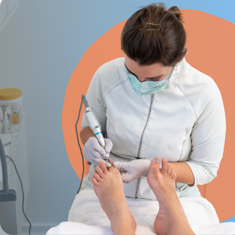
Premier Foot & Ankle delivers expert diagnosis and treatment for a full range of foot and ankle conditions—from everyday care to advanced injuries and surgical needs. Our award-winning team proudly serves Venice, Florida, with personalized, compassionate care focused on your comfort and long-term wellness. Because when it comes to your feet, only the best will do.
Conditions We Treat
Premier Foot & Ankle delivers expert diagnosis and treatment for a full range of foot and ankle conditions—from everyday care to advanced injuries and surgical needs. Our award-winning team proudly serves Venice, Florida, with personalized, compassionate care focused on your comfort and long-term wellness. Because when it comes to your feet, only the best will do.
Meet Our team
Get to know the compassionate experts dedicated to keeping you on your feet—meet the team behind Premier Foot & Ankle Specialists.
Dr. Brielle Roggow
DPM
Dr. Brielle Roggow was born and raised in Jackson, Minnesota. She attended and graduated from Minnesota State University, Mankato, with a bachelors of science in biology.
Read More
Dr. Jeremy Bonjorno
DPM
Dr. Bonjorno is a community-focused podiatrist with a commitment to high-quality patient care. His podiatric interests include diabetic foot care, wound care, bunions, hammertoes, neuromas, and plantar fasciitis.
Read More
Coming Soon
Amy McCreery
Office Manager
Amy McCreery is the dedicated office manager at Premier Foot & Ankle Specialists, known for her warm personality and attention to detail. She ensures every patient has a smooth and stress-free experience from check-in to follow-up.

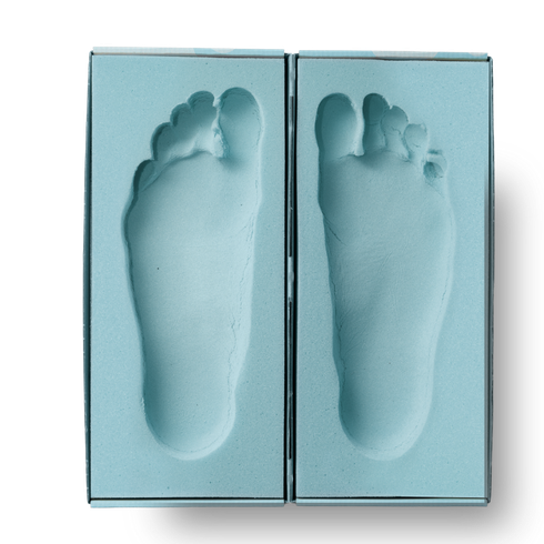
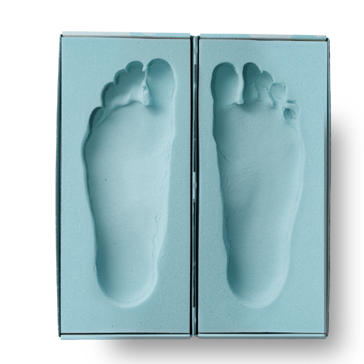
Custom Orthotics provide the Comfort and Support Where You Need It Most
Custom orthotics can make a world of difference in your daily comfort. We design prescription orthotics to relieve pain, improve stability, and support conditions like arthritis, flat feet, and heel pain. Our experienced team will assess your needs and create orthotics that fit your lifestyle—so every step feels easier and more supported.
Schedule your new patient appointment today and take the first step toward lasting relief.


